Background: Lymphocytic-plasmacytic enterocolitis (LPE) is a rare, poorly understood form of infiltrative intestinal disease in horses, with a generally grave prognosis. Corticosteroids are the drugs most commonly used in treatment, but there are currently no clinical guidelines in terms of dose rates and duration of treatment, and systemic administration can cause significant adverse effects, particularly in animals with comorbidities (Vitale 2022).
Objectives: To describe the successful treatment of two cases of LPE with differing presentations that underwent thorough diagnostic evaluation and failed to respond to conventional therapy. The importance of addressing the gut microbiome- immune system axis is emphasized.
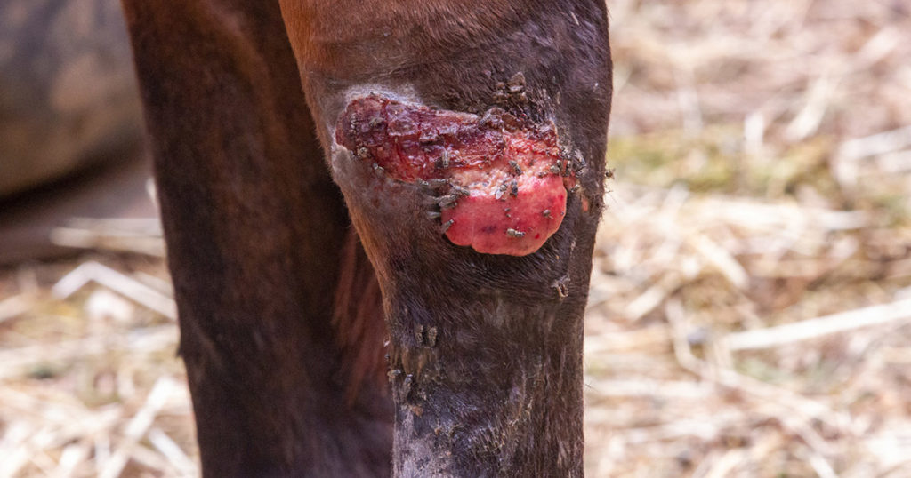
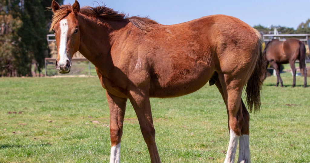
Study design and data collection: Retrospective analysis of medical records. Case details and setting: Case one: a 10-year-old Quarter Horse gelding with diffuse LPE confirmed by laparoscopic biopsy. Case two: a 12-year-old miniature gelding with diffuse LPE diagnosed by intestinal biopsies and transabdominal ultrasonography. Both horses had undergone repeated extensive physical examinations, gastroscopy (with no lesions evident) as well as complete blood counts and basic metabolic panels. Case two had undergone faecal egg counts and microbial culture. Both horses were resident in Australia. Case one presented with a 15-week history of lethargy and depression, ventral oedema extending from the jaw to the prepuce and daily episodes of mild to severe colic. Colic events were managed with per os administration of flunixin meglumine, but not always successfully. Daily medication consisted of oral dexamethasone, omeprazole and vegetable oil. The horse was also receiving a proprietary GI- specific dietary supplement and had previously been treated with sucralfate. Complications with wound healing following the laparoscopy had necessitated an extended period of hospitalization. Case two presented with sudden onset, profuse watery diarrhoea of 2 years ‘duration; bruxism; intermittent passing of mucus; lethargy and sporadic inappetence for hard feed. At the time of assessment, 8 weeks after LPE diagnosis, the gelding was receiving daily oral doses of dexamethasone, omeprazole and codeine. Previous treatments included a five-day course of benzimidazole followed by single doses of moxidectin and praziquantel; bentonite; yoghurt; oral faecal transplantation; misoprostol; corn oil; psyllium husk; phenylbutazone; sucralfate and antimicrobials (metronidazole and sulfadiazine/trimethoprim).
Intervention: Case one received treatment with an ethanol-based preparation of astragalus, echinacea, chamomile, cramp bark, ginger and liquorice in combination with glutamine, zinc citrate, vitamin D3, slippery elm bark and a multi-strain probiotic. The vegetable oil was stopped immediately. A compound of corydalis, California poppy, valerian and cramp bark was used to manage colic episodes, as required. After 1week, the gelding was weaned off the dexamethasone and omeprazole. The diet was modified to increase dry matter and hard feed meal sizes were reduced while the horse was transitioned to a grain-free diet. Walking exercise was introduced and the owner was encouraged to leave the horse without rugs/ blankets. Case two was administered an ethanol-based blend of goldenseal, meadowsweet, echinacea, corydalis, aniseed and ginger alongside glutamine, zinc citrate, vitamin D3, slippery elm bark and a multi-strain probiotic. Concentrate feed and oaten chaff were withheld temporarily, with lucerne and teff hay quantities increased. Dexamethasone, omeprazole and codeine were gradually curtailed while hand walking was introduced and the pony was left without rugs/blankets.
Results: Case one: 24- h video surveillance confirmed significant reduction in severity and frequency of colic episodes within 2 weeks. The ventral oedema resolved after 4 weeks, with noticeable improvements in perceived energy levels. The gelding has had no recurrence of oedema for 20 months and has been colic-free for 14 months. Competition use has resumed. Case two: within 3 weeks bruxism had ceased, there was a notable improvement in manure density and markedly improved energy levels. After 3 months, faecal output was normal on most days and a grain-free balancer feed and lucerne chaff were introduced, which the pony readily consumed. By 5 months manure density was normal, and has remained so after 2 years.
Main limitations: Biopsies were not repeated after treatment and recovery; efficacy of treatment based on observation of clinical signs; small case numbers.
Conclusion: Chronic LPE, including cases unresponsive to pharmaceutical intervention, can be stabilized and treated successfully by integrating nutraceutical, herbal medicine, dietary and management strategies.
Ethical animal research: No ethical approval required.
Funding information: None.
Conflict of interest statement: None.
Reference:
Vitale, V. (2022) Inflammatory bowel diseases in horses: What do we know? Equine Vet. Educ. 34, 493–500.
Published in Equine Veterinary Education Volume 36 | Supplement 13 – Link
Camilla Whishaw is a highly regarded, experienced horsewoman and naturopath, helping to holistically treat and manage a broad range of equine health conditions and injuries, with a passion for mare and stallion fertility.
As a world-renowned practitioner, presenter, author, and consultant in the field of Equine Naturopathy, Camilla shares her knowledge through keynote presentations, interviews, lectures, panel sessions, and workshop training.